
An investigation by University of Michigan researchers reveals new insight into why shoulder injuries take a long time to heal.
Specifically, the researchers focused on why rotator cuff injuries tend to accumulate fatty tissue instead of new muscle.
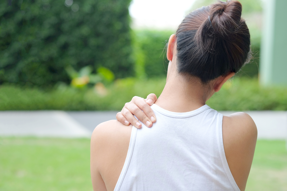
Tears of the rotator cuff, the group of four muscles and tendons that help to stabilize the shoulder during movement, are very common.
“Rotator cuff injuries happen fairly frequently, and the majority of them are as a result of regular wear and tear caused by aging,” said Dr. Bill Johnson, a Dallas, Texas, stem cell physician.
Injuries develop as tiny tears in the muscle tissue that get bigger over time.
“Symptoms start as nagging pain and stiffness, then eventually progress to constant pain and loss of function,” Johnson said.
Treatment for rotator cuff injury includes self-care steps such as ice, heat and rest. Some individuals see success with nonsteroidal anti-inflammatories (NSAIDS) like ibuprofen or naproxen.
Advanced cases may require steroid injections or even surgery if the injury is severe enough.
Surgical repair of the rotator cuff means weeks of recovery and keeping the surgery immobile. After surgery, there are no guarantees that the joint will be as good as new.
“After surgery for rotator cuff injuries, some patients experience muscle weakness and atrophy,” Johnson said.
One reason for this could be that rotator cuff injuries tend to develop fat accumulation at injury sites. Fat development after rotator cuff injuries occurs more frequently than in any other injured muscles.
The Michigan scientists wanted to find out why this happens.
To answer the question, they took a deep dive into the cellular, molecular and genetic reasons behind the development of fat.
Using mice, the study authors removed specific stem cells known as satellite cells from both the rotator cuff muscle and the calf muscle.
Although the muscle stem cells were generally thought to be the same, both types of satellite cells were investigated to examine gene expression to identify if any differences existed.
And they did.
After analyzing the two types of stem cells, the researchers found that those from the rotator cuff muscle behaved differently and also had different genetic markers.
The rotator cuff stem cells differentiated into muscle cells, but 23 percent less than those taken from the calf.
The rotator cuff stem cells differentiated into 23 percent fewer muscle cells than the calf muscle stem cells. Rotator cuff cells also had an 87 percent decrease in muscle formation than calf muscle stem cells.
What the rotator cuff stem cells did have were more genetic markers that caused fat cells to generate – up to 65 percent more than calf muscle stem cells.
The next step was identifying why the genetic markers caused fat cell production to become activated instead of muscle cells.
Upon further analysis, the researchers found that the rotator cuff muscle stem cells had 355 regions of DNA that were different from the stem cells taken from the calf muscle.
Using a pathway-enrichment analysis, they were able to identify what genes triggered the development of fat, a process known as adipogenesis.
The results of their investigation revealed that rotator cuff muscle stem cells contain DNA that makes them more apt to develop into fat cells.
The Michigan study is the first to study the impact of DNA and how it can hinder healing for rotator cuff injuries. The researchers hope that their investigation is the first of many studies about fat and muscle healing and develop new treatments for treat musculoskeletal conditions.
The premise is exciting for Johnson.
“Learning about the things that can hinder healing can help develop workarounds or new approaches to treating patients,” Johnson said.
Source: Michigan Medicine – University of Michigan. “Stem cells provide greater insight into rotator cuff disease.” ScienceDaily. ScienceDaily, 26 February 2019.